A. Alonso Aguirre, Colin J. Limpus, Terry R. Spraker, and George H. Balazs
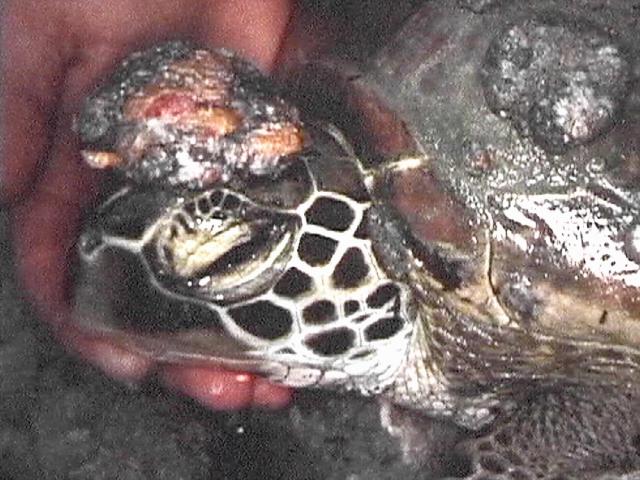
Photo by Colin Limpus
A survey took place in Moreton Bay, Australia in mid-June 1998. Multiple sites in Moreton Bay were selected for examination of turtles. These turtles were captured by "rodeo technique" and brought to a research vessel and were then evaluated for disease processes and health status. Turtles were examined, measured and blood samples were taken and all their orifices were checked carefully. Tumors were biopsied, some frozen, some submitted for histopathology.
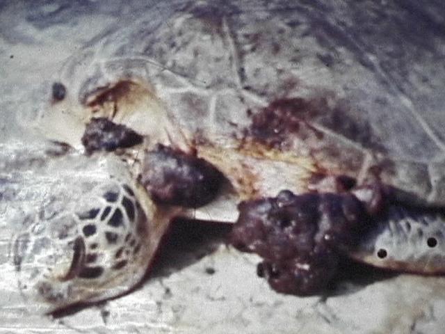
Over a week's span, the research group caught 120 green sea turtles and 50 loggerheads. Of the green sea turtles, 17% had fibropapillomas and 8% of the loggerheads had FP. The fibropapillomas ranged from very small to 10-15 cm.
The tumors were frozen and searched for viruses, especially herpesviruses. The tumors were also taken and put in formalin and examined.
Dr. Spraker then details the results of those biopsies comparing Australian fibropapilloma tumors with those from Hawaii, Costa Rica and Mexico.
Australian samples showed more inflammation in the normal skin sections and a greater abundance of bacteria than in the normal skin samples of other populations.
The tumors of Australia and the tumors from Hawaii look very much the same to a point of being barely distinguishable. The tumors from Mexico and Costa Rica look similar except for one thing: there is much more of a cell-mediated response in the dermis of Mexican and Costa Rican samples. Cell-mediated immunity in the Costan Rican and Mexican samples was about 40%-50%, compared to those of Hawaii with just 2%. This type of cell immunity can lead to regression.
Peter Bennett1 (honu@turtles.org), Ursula Keuper-Bennett1 (howzit@turtles.org) and George H. Balazs2 (gbalazs@honlab.nmfs.hawaii.edu)
1Turtle Trax, 24 Reid Dr, Unit 3, Mississauga, Ontario, Canada L5M 3A6
2National Marine Fisheries Service, Southwest Fisheries Science Center, Honolulu Laboratory, Honolulu, 2570 Dole Street, Honolulu, Hawaii 96822, USA
My wife, Ursula, and I spend each July and August diving in the waters of Honokowai, West Maui, Hawaii. She's a teacher and I'm a technical writer, and we spend most of our dives watching, photographing, and videotaping Hawaiian green turtles, or honu. We can do this because the honu are among the tamest of nature's wild creatures.
We started in 1988, when we met Clothahump, our first sea turtle. By 1990, we had explored Reef 2 and discovered the Turtle House, the two places where turtles congregate at Honokowai. Many of the turtles we saw had fibropapilloma (FP), so we reported this to George Balazs of the NMFS Honolulu Laboratory. George provided us with background information about the disease. From the material he supplied, this quote from Herbst (1994) stood out:
"The number of turtles that develop GTFP in the wild over time (incidence) and the proportions of affected turtles that develop severe disease and die (morbidity and mortality) are unknown. These data are desperately needed if we are to understand the demographic impact of GTFP on wild populations."
We were concerned about the turtles. We wanted to do something. This quote spoke to us because it meant that by turtle-watching, we might be able to help.
We went on to take nearly 4000 pictures and over 150 hours of videotape. In 1997, George reminded us that it was time to quantify things, so we built a database. Then, with George's guidance and help, we set out to analyze this material.
To tell the turtles apart, we relied on the patterns formed by the scales on their faces--their profiles. This pattern allows us to identify individuals from year to year. For each individual, we recorded their profiles, their sexual maturity, and if present, their tag information.
For every year that we saw a turtle, we also recorded a count of tumors by size for external surfaces of the turtle, an overall estimate of the prevalence and severity of FP, and an estimate of the size of the turtle.
To count and evaluate tumors, we used the method developed by Balazs (1991) with the help of John Sundberg, and later refined by Work & Balazs (this volume). This system places tumors in four sizes: A is anything less than 1 cm in diameter, B includes tumors from 1 to 5 cm, C tumors are from 5 to 10 cm, and D includes anything over 10 cm.
The method also includes an overall score for the turtle. This is a subjective estimate based on the total number and size of tumors. There are three categories for tumored turtles: light, moderate, and heavy.
We were keen to assess the impact of FP on juvenile turtles. To do this, we followed size assignments used by Balazs for honu since 1973: juvenile (post-pelagic up to 65 cm), sub-adult (65 up to 85 cm), and adult (larger than 85 cm).
Although we don't take measurements in the wild, photos do let us make estimates by comparison with objects of known size.
Because our data are gathered from images, it isn't possible to get a complete assessment for an animal. For example, we can't check whether a turtle has tumors deep inside the mouth. Sometimes we have a record of only part of a turtle. Our data, therefore, are skewed in a manner that underestimates the occurrence of FP.
We define regression as follows: if a tumor has gotten smaller or become undetectable in our photos and video, we call that regression. We first noticed regression in Tutu, who had a size B eye tumor in 1990. By 1993, however, her eye tumor had almost disappeared. Finally, our photos from 1997 leave no doubt that the tumor has vanished. Since 1996, we've seen more and more animals that show some regression. The question, therefore, was how many tumored Honokowai turtles were regressing?
We identified 247 turtles. We found that 64% (n=158) had FP at some point. We have seen 37% (n=91) of the 247 in more than one summer. We consider these turtles to be Honokowai residents, and we call them "resights." Of our 91 resights, 66 or 73% have had FP, and from our 66 resighted turtles with FP, we have photo evidence of regression for 32%, or 21 individuals.
Of these 21 regressed animals, we have presumed 52% (n=11) are sexually mature. Three are females that were tagged while nesting at the French Frigate Shoals. The other eight are males, judged by the fact that their tails have grown beyond their hind flippers. As judged by size, 17% (n=43) out of 247 turtles were classed as juveniles at some point in our records. 60% (n=26) of these 43 had FP. Nineteen of the juvenile turtles have been resighted. So far, 74% (n=14) of them have eventually developed FP. We have documented regression for only one resighted juvenile.
Course of the disease: In 81% (17 out of 21) regression cases, we saw the turtle get worse before it got better. Infected turtles had light tumors at the onset, got visibly worse in the second summer, and peaked in the third. In turtles that showed regression, tumor growth either stabilized or reversed itself after the third summer. This was usually followed by two summers of steady regression, after which tumors often became undetectable by examination of our images. We have documented this level of regression in 11 cases, all animals that had A and B sized tumors only.
This leaves 10 cases in which the tumors can still be seen. In 7 of these cases, regression is still underway. The other three cases had tumors that reached size C, however. While these C-sized tumors have regressed significantly, it appears that they might not disappear completely.
Tumor scoring is a reliable indicator: The subjective tumor score does help to predict outcomes. Of the 66 resighted turtles that have had FP:
Moderately afflicted adults can recover: Tiamat is the most severely afflicted adult that we've seen regress. We've known her since 1992, when she was fine. For 1994, however, we classed her as moderately afflicted. Unexpectedly, by 1995 her condition had improved. In 1996, we happily added her to our list of regression cases. We didn't see her in 97, but in 98 she bore an engraved V40 on the right side of her carapace--proof that she had nested at the French Frigate Shoals in ‘97.
Only one juvenile has ever regressed: Another moderate case is our most interesting and remarkable story. Kamaha'o is Hawaiian for "remarkable." This is the only juvenile turtle that we have ever seen regress. In 1994, Kamaha'o a had eye and mouth tumors, and was notably emaciated. This turtle looked to be doomed. In fact, we're so accustomed to youngsters disappearing on us that we didn't immediately recognize this robust turtle, photographed in 1997, as the tumored little beast from 1994. It was only in the preparation of this paper that we made the connection. This showed us that FP does not have to be a death sentence for juveniles.
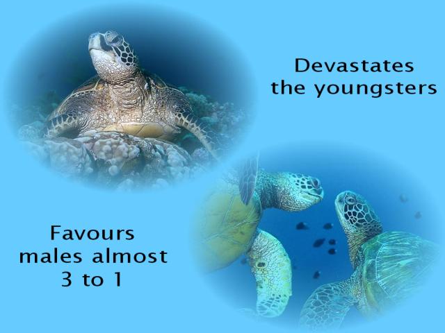
Juveniles affected most severely: Nevertheless, our data show that regression clearly favours larger turtles. To recap: only 5% or 1 of 21 regression cases was a juvenile (based on size), while 21% or 14 of the 66 resighted turtles that have had FP were juveniles at some point. While our sample is small, this hint that FP cuts a deep swath through the little ones is echoed in data from Kaneohe Bay, Oahu, Hawaii. There, most turtles sampled are juveniles and the FP regression rate is only 4.5% (Balazs et al. In press).
Males vs. females: At first, the regression rate for subadult and adult turtles seems encouraging. 38% (n = 20) of 52 larger resighted turtles that have had FP have regressed. It's instructive, however, to look at the 11 sexually mature turtles that we identified in the regression cases: eight of them are male. If our data are typical, almost three times more males are recovering than females. This reflects work done by Koga and Balazs (1996), who report significantly higher FP in female honu based on necropsies of hundreds of stranded animals.
Potential population impact: Since we have no other data to compare, we don't know if what we are seeing is typical of the broader FP picture. We hope it isn't, but if it is, we have a disease that:
The implications for the honu are troublesome.
Environmental concerns/potential for future study: Recent findings by Landsberg et al. (In press) have suggested there is preliminary and provocative evidence linking high concentrations of dinoflagellates (specifically Prorocentrum) to a high prevalence of fibropapilloma. One of the sites they sampled for these organisms was Honokowai.
Early findings suggest the honu might be suffering from chronic exposure to okadaic acid produced by the dinoflagellates living in the seaweeds that the honu use for food. It is difficult to watch turtles eat when we know it is possible that they are slowly poisoning themselves. Still, we continue to document their foraging habits. For one thing, we hope to understand why the disease affects the little ones so profoundly.
Of course, if FP is indeed fueled by eutrophication, which in turn is caused by run-off, sewage, animal wastes, and the destruction of wetlands, we are forced to wonder about the future of any animal that is dependent on clean water.
Finally, as if the current state of the coastal waters isn't worrisome enough, a new storm cloud blew into Honokowai last summer. Tutu (Hawaiian for grandmother), known since 1990 and our first regression case, showed troubling white anomalies on her neck and shoulders.
While this might be the beginning of a dose of a new type of barnacle we've been seeing on the turtles only recently, or perhaps something else entirely, we cannot rule out the possibility that this could be the harbinger of the return of FP.
In summary, we have collected a considerable body of photo documentation of the prevalence of FP in a community of Hawaiian turtles. Through retrospective examination of this data, we have shown that about one in three diseased turtles has regressed, but a closer look at exactly which turtles regress reveals disturbing patterns: juveniles rarely regress, and recovering males outnumber females three to one. Finally, there is one last sobering thought--Tutu's condition has raised an ugly, troubling question: is regression permanent?
Dr. Larry Herbst, Richie Moretti, and Dr. Grahame Webb for their encouragement and support.
Balazs, G. H. 1991. Current status of fibropapillomas in the Hawaiian green turtle, Chelonia mydas. In G. H. Balazs and S. G. Pooley (eds.), Research plan for marine turtle fibropapilloma, p. 47-57. U.S. Dep. Commer., NOAA Tech. Memo. NMFS-SWFSC-156.
Balazs, G. H., S. K. K. Murakawa, D. M. Ellis, and A. A. Aguirre. In Press. Manifestation of fibropapillomatosis and growth rates of green turtles in Kaneohe Bay, Hawaii. In: Proceedings of the Eighteenth Annual Symposium on Sea Turtle Biology and Conservation, March 3-7, 1998, Mazatlan, Mexico. U.S. Dep. Commer. NOAA Tech. Memo. NMFS-SEFSC.
Herbst, L. H. 1994. Fibropapillomatosis of marine turtles. Annu. Rev. Fish Dis. 4:389-425.
Koga, S. K. and G. H. Balazs. 1996. Sex ratios of green turtles stranded in the Hawaiian Islands. In J. A. Keinath, D. E. Barnard, J. A. Musick, and B. A. Bell (comps.), Proceedings of the Fifteenth Annual Symposium on Sea Turtle Biology and Conservation, February 20-25, 1995, Hilton Head, South Carolina, p. 148-152. U.S. Dep. Commer. NOAA Tech. Memo. NMFS-SEFSC-387.
Landsberg, J. H., G. H. Balazs, K. A. Steidinger, D. G. Baden, T.M. Work and D. J. Russell. In press. The potential role of natural tumor promoters in marine turtle fibropapillomatosis. Journal of Aquatic Animal Health.
Work, T. M. and G. H. Balazs. This volume. Quantification of tumor severity and hematology in green turtles afflicted with fibropapillomatosis in the Hawaiian Islands.
Félix Moncada G. and Adela Prieto T.
Transcribed from the translation provided at the Symposium by Jeff Seminoff. Photo by Elvira Carillo.
In Cuba, fibropapilloma (FP) has been seen since the middle of the 1980's with fishermen reporting the disease. Aims of the current Cuban study are to determine prevalence of this disease on the Cuban shelf and ultimately to contribute to the knowledge of this disease and its treatment.
Between 1984 and 1996 the surveys were carried out from the 14 commercial fisheries along the Cuban shelf. In total, 3390 green turtles were sampled from this region. When an animal was captured, straight carapace length/width, weight, as well as location and date of capture were recorded.
 In case of infected animals, the presence and size of tumors were recorded.
Of the 3390 green turtles sampled, 20 or 0.006% were infected with fibropapilloma. The first case was documented in 1985 but caution that no sampling was done prior to 1985. Of the turtles infected the size of tumors were measured for seven. The tumors varied between 3 and 22 cm. The straight carapace length of these seven ranged from 55 and 102 cm with average of 77.5 cm.
The highest percentage was found in females at 90%. [Note: This bias reflects the sex ratio of animals recorded in the harvest, around 80% female.]
The infected animals were detected in movement corridors along the north eastern region of Cuba, as well as in off shore areas near reproductive zones/nesting beaches and finally along feeding grounds to the south.
The greatest amount of green turtles were detected in 1985 when ten animals were seen. One or fewer cases from 1990 onward. However this does not mean that the disease has decreased or kept steady. Some of the fishermen have seen some indications of little to medium sized turtles with tumors or warts in many of the feeding areas.
Since 1996 there have been only two capture sites with a minimum quota of animals captured thus the probability of finding individuals having fibropapillomas throughout the Cuban shelf has decreased. I would like to add from the prior statement that only the largest animals are captured in the fisheries so all the small animals that might have fibropapillomas are not sampled.
In Cuban waters, like other areas of the Caribbean, fibropapillomas do occur, however only occasionally. Studies have been started recently to investigate other areas on the Cuban shelf which will probably increase the probability of finding this disease in other areas.
 Sickbay
 Table of Contents