 Left eye, 1995 Turtle 24: Nani (64K JPEG)
We offer these images to anyone who can make use of them. We hope that someone can shed more light on what exactly these photos reveal about the eyes of these turtles.
In the Honokowai population, fibropapilloma tumors almost always begin in the eyes, and without exception the tumors appear first in the posterior corner of the eye. If the turtle has tumors in both corners of an eye, the posterior tumor is always larger. Eye tumors often cause drastically reduced vision or blindness.
These images were originally taken underwater at Honokowai, Maui, during the summers of 1994 and 1995. This is not macro-photography. These images have been prepared by loading the highest resolution CD-ROM image possible, then cropping only the eye. At times, this means less than ideal sharpness.
If the original image from which a close-up was taken is not online and you want to see it, ask webmaster@turtles.org.
The icons below are linked to 640x480 JPEGs. If you prefer to review the images as 240x180 GIFs, use our contact sheet (9 GIFs, 201K).
The nature of this medium makes it impossible for us to know exactly how our images will appear on other monitors. Colours can be shifted or missing entirely. The brightness, contrast, colour balance, and gamma of our monitors can vary significantly from yours. This means that you must not rely on the colours contained in these images, even if you are using a video system that has millions of colours.
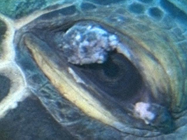
When we originally posted this image we believed it to be a healthy eye, based on the advice of one expert. Another expert has since said the eye could be showing the earliest signs of tumor growth. We have therefore removed it as an example of a healthy eye.
Unfortunately, if we cannot use Aikane, we do not have an example of a healthy eye.
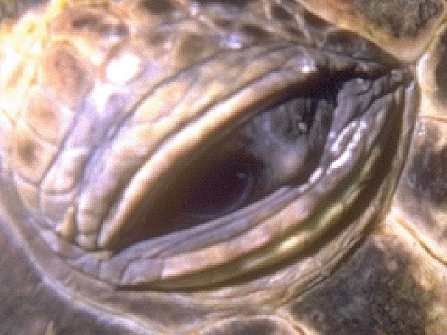
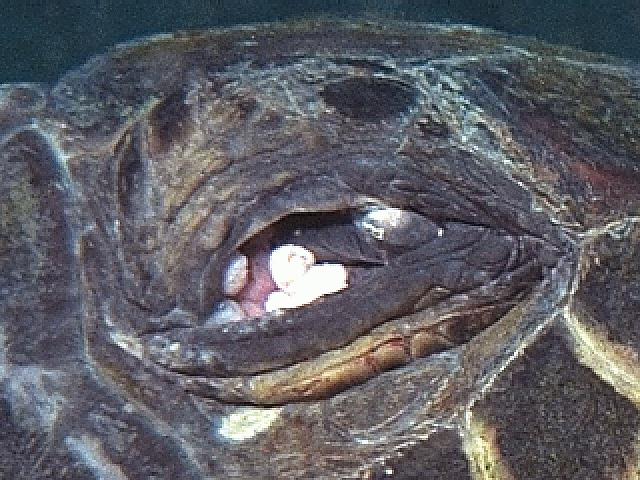
Left eye, 1995 Turtle 24: Nani (64K JPEG)This is the best example we have of early tumor growth. This turtle also has the salt and pepper spotting that we normally see develop into tumors.
 Left eye, 1992 Turtle 3: Howzit (69K JPEG)
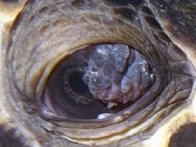
This tumor is no more than eleven months old. When we left at the end of the summer of 1994, Howzit's eyes did not have visible tumors, although we suspected they were beginning to grow.
 Right eye, 1993 Turtle 27: Hilu (45K JPEG)
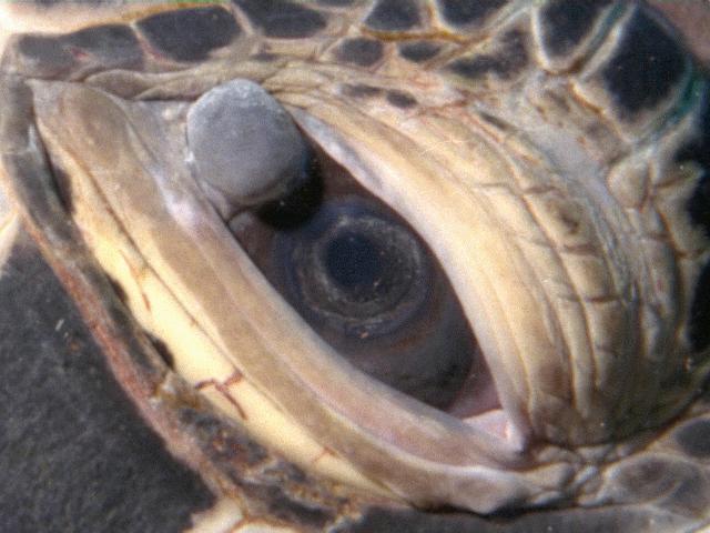
This is an example of a tumor that is in regression. Hilu's right eye has cleared significantly since 1994. Roughly 15% of the Honokowai regulars have shown some signs of remission.
 Left eye, 1995 unidentified (64K JPEG)
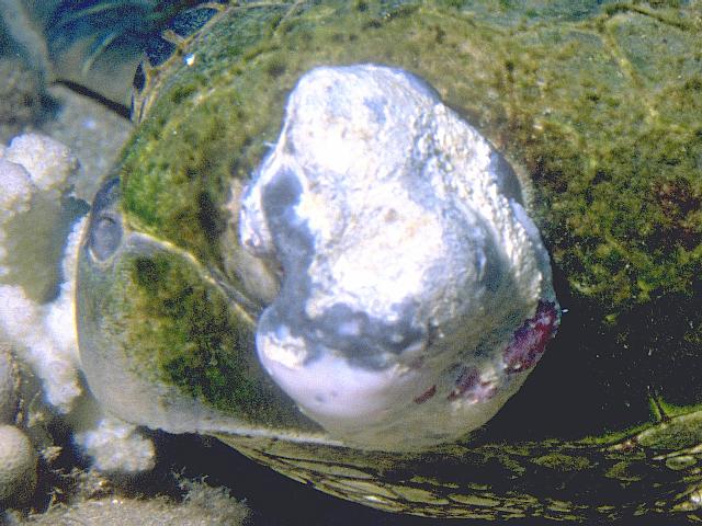
This is an extreme example of an eye tumor. We could not determine whether this turtle is in our logs because the combination of tumor and algae completely covers the profile.
 Left eye, 1991 Turtle 10: [unnamed] (76K JPEG)
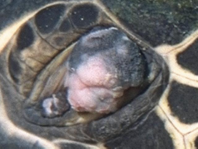
In 1992, this turtle had a tumor that completely obscured the left eye. Now the eye is gone, but we have no idea why. We are not sure exactly what is occupying the eye socket.
 Right eye, 1992 Turtle 26A: Raphael (45K JPEG)
This is an example of a double tumor. In 1992, Raphael showed no visible signs of tumors. In 1993, we suspected tumors were starting in the corners of each eye. This image is from 1995.
 Left eye, 1995 Turtle 38: [unnamed] (53K JPEG)
Aside from exhibiting the usual tumors in both corners of the eye, this turtle has unsual growth surrounding the eye. This extends across the face and into the nostrils. We have not seen this in any other case.
 Left eye, 1992 Turtle 22: 4 Spot (43K JPEG)
4 Spot was a clean juvenile upon arrival at the Turtle House in 1992. By 1995, a double tumor had almost covered the left eye.
If there are specific images you would like to see in the Turtle Trax pathology pages, let us know. Please send comments or suggestions to webmaster@turtles.org.
 Sickbay
 Turtle Trax Home Page