 Polzbarney [1995 Turtle 15] (63K JPEG)
We offer these images to anyone who can make use of them. We hope that someone can shed more light on what exactly these photos reveal about the tumors of these turtles.
These images were originally taken underwater at Honokowai, Maui, during the summer of 1998.
The icons below are linked to 640x480 JPEGs. If you prefer to review the images as 240x180 GIFs, use our contact sheet (9 GIFs, 324K).
The nature of this medium makes it impossible for us to know exactly how our images will appear on other monitors. Colours can be shifted or missing entirely. The brightness, contrast, colour balance, and gamma of our monitors can vary significantly from yours. This means that you must not rely on the colours contained in these images, even if you are using a video system that has millions of colours.
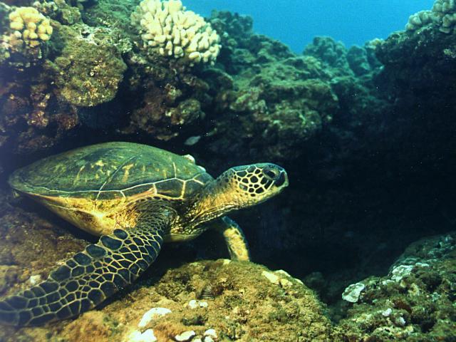
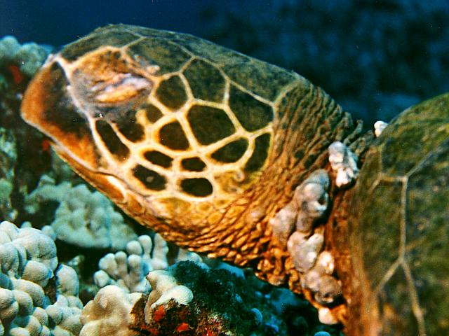
Polzbarney [1995 Turtle 15] (63K JPEG)Polzbarney, under his home ledge in Summer 1998. He's now lived in this same location since 1995, demonstrating the remarkable site fidelity of Hawaiian green sea turtles. Compare the barely visible growths on the right shoulder in 1997 with the tumor there just 10 month later.
 Polzbarney [1995 Turtle 15] (64K JPEG)
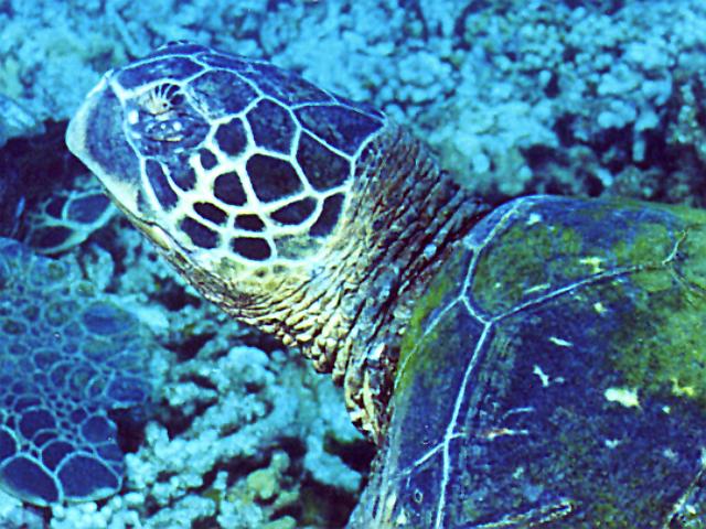
Polzbarney, under his home ledge in Summer 1997. The first eruptive tumors are evident on the eyes, and somewhat less so on the right shoulder, which has a cluster of white pimple-like growths.
 Unnamed [1998 Turtle 44] (61K JPEG)
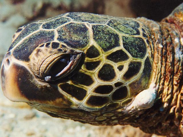
1998 Turtle #44, close up left profile. These are typical beginning eye tumors. White blemishes on the neck often herald the development of a "tumor collar." Most unusual is the tumor growing out from between facial plates, which is also the largest tumor this turtle presently has.
 Unnamed [1998 Turtle 70] (60K JPEG)
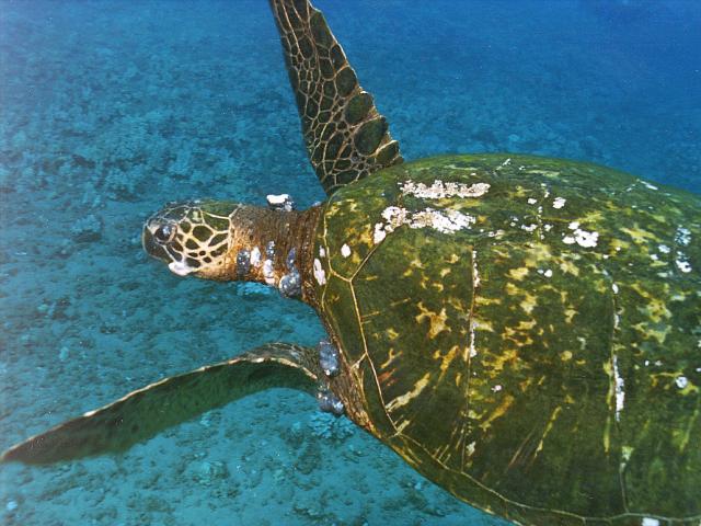
Turtle 1998 #70, photographed in 1997. Note the mouth tumor and multiple growths over neck and shoulders.
 Unnamed [1998 Turtle 70] (61K JPEG)
1998 #70 photographed this year. The increase in tumor growth is notable.
 Unnamed [1998 Turtle 70] (72K JPEG)
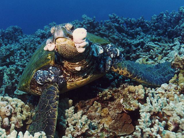
Turtle 1998 #70 has worsened considerably since the previous summer. Mouth tumors have ballooned and threaten to block breathing and feeding.
 Unidentified [Unlogged] (51K JPEG)
As yet unidentified/unlogged turtle photographed at the end of August 1998. Aside from the many tumors burdening this turtle (the most life-threatening is the mouth tumor) the white cuts and gashes to its carapace announce its recent face-to-face with a large shark, probably a tiger. The numerous scratches are most likely the result of the shark trying to get a "perch", a bite-hold on the turtle, and not penetrating far enough to hold tight. Our shark expert suggested that some shaking was involved because that's how tigers "saw" through large tough animals.
 Unnamed [1994 Turtle 12] (60K JPEG)
Example of regression. This is the left profile of 1994 Turtle #12 in 1996. Note the loss of the left eye to fibropapilloma disease. The turtle is also burdened with numerous growths over neck and shoulders. In 1996, we assumed this animal was a goner.
 [1994 Turtle 12] (70K JPEG)
Here is the same turtle two years later, in the Summer of 1998. Only close examination could detect the darkened and shrunken tumors The left eye is now just a slit (we assume the eyeball was eaten away by disease and parasites) but otherwise this turtle's condition has improved immensely, and so can be added to our growing list of regression cases.
If there are specific images you would like to see in the Turtle Trax pathology pages, let us know. Please send comments or suggestions to webmaster@turtles.org.
 Sickbay
 Turtle Trax Home Page