Drs. Larry Herbst and Paul Klein organize their slides for their talks. (59K JPG)
A. Alonso Aguirre1
| 1Joint Institute for Marine and Atmospheric Research, 2570 Dole Street, Honolulu, Hawaii 96822-2396, USA |
As a request to the National Marine Fisheries Service (NMFS) Honolulu Laboratory by the organisers of the 18th Annual Symposium on Sea Turtle Biology and Conservation, held in Mazatlan, Sinaloa, Mexico, 3-7 March 1998, Alonso Aguirre was asked to coordinate a Fibropapilloma Workshop on 5 March. The following constitutes a report requested by the editors of the Marine Turtle Newsletter/Noticiero de Tortugas Marinas.

| Drs. Larry Herbst and Paul Klein organize their slides for their talks. (59K JPG) |
The three-hour meeting had as a primary objective to update sea turtle biologists on current research, recent findings, and specifically, the advances regarding the identification and characterisation of possible aetiological agents associated with the condition. This well attended workshop consisted of a panel of scientists who gave 10-15 minute presentations on their current research, followed by a stimulating question and answer period.
Fibropapillomatosis (FP) is a disease characterised by multiple cutaneous tumor masses ranging from 0.1 to more than 30 cm in diameter that, to date, has primarily affected green turtles (Chelonia mydas). In addition, the condition has been reported in other species including loggerhead (Caretta caretta), olive ridley (Lepidochelys olivacea), Kemp's ridley (L. kempii) and flatback (Natator depressus) turtles. However, of these species, FP has only been histologically confirmed in loggerhead turtles from Florida and olive ridley turtles from Costa Rica. The disease has a world-wide circumtropical distribution and has been observed in all major oceans. Where present, prevalence of the disease varies among locations, ranging from as low as 1.4% to as high as 90%. Although several viruses have been identified associated with the tumors, the primary aetiological agent remains to be isolated and identified.
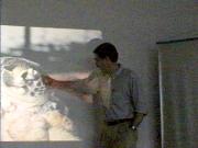
Dr. Larry Herbst points to ocular tumors on a Florida green sea turtle. (47K JPG) | 
|
Larry Herbst (Albert Einstein University, New York, USA) provided an overview of FP. The two main aetiological hypotheses were discussed: The non-tumor hypothesis supports the theory that these growths may be a cellular reaction to scar tissue or may represent hyperplastic tissue. The tumor hypothesis regards these growths as neoplasia caused by any of a number of different agents including chemicals. Several transmissible agents have been postulated including parasite eggs (spirorchid trematodes or blood flukes), bacteria and viruses. Two groups of viruses have been identified as associated with the tumors: The enveloped viruses which are sensitive to environmental factors, with herpesvirus and retroviruses belonging to this group. The non-enveloped viruses are environmentally resistant and examples include papillomaivurs and polyomavirus. A herpesvirus has been identified in >95% of the tumors studied in Florida. This virus was not found in normal turtle skin or other normal tissues. Green and loggerhead turtles from various locations in Florida have been diagnosed with FP and Herbst's transmission studies have demonstrated the involvement of a filterable, subcellular agent of infectious nature. Direct experimental inoculation of turtles has consistently demonstrated a horizontal transmission and tumors have developed at the site of inoculation in turtles free of the disease within a year post-inoculation.

| Lew Ehrhart gives a talk on the Indian River during the FP workshop. (54K JPG) |
Lhewellyn Ehrhart (University of Central Florida, Orlando, USA) has monitored green turtles during stunning events caused by low water temperatures along east central Florida at the Mosquito Lagoon since 1975. His long term studies have shown that during the 1970's there was no evidence of the disease; however, few animals were handled at the time. By 1982, the studies shifted to the central region of the Indian River system, about 120 km south of Mosquito Lagoon. Turtles with FP were identified immediately with an annual prevalence since 1982 of 28-65%. During 1985, FP was found in 29% of 145 turtles stunned at Mosquito Lagoon. During 1996-97, 61-63% of the turtles examined presented FP and during 1998, 82% of the turtles had tumors. At another field site off Indian County, no FP was observed prior to 1996. Logistic difficulties in netting animals, poor underwater clarity and algae-covered reef structure were present; however, during 1997 about 20% of turtles presented FP. Ehrhart's research suggests that green turtles free of FP migrate to the neritic zone along the Florida coast and remain free of tumors while residing there. Many of these turtles may become infected when they enter the lagoon system.

| George Balazs gestures during his presentation at the FP workshop. (49K JPG) |
George Balazs (NMFS Honolulu Laboratory, Hawaii, USA) provided an overview of the Hawaiian situation with emphasis on strandings and manifestation of FP in the foraging pastures of Kaneohe Bay, Oahu; Palaau, Molokai; and the entire west coast of the island of Hawaii (where the disease is absent). Balazs, with other members of NMFS Honolulu Laboratory, convened a FP workshop in Honolulu in 1990. The Research Plan produced as a result (Balazs & Pooley 1991) has been an important source reference for all FP researchers. More recently, another fibropapilloma workshop was held in 1997 by NMFS in Honolulu to provide an update of the significant advances made with FP research. NMFS perspective and interest in the disease are based on several concerns, as follows: green turtles in Hawaii are protected under the Endangered Species Act and are classified as Threatened; the possible role of toxic marine pollutants in causing tumors; the role of sea turtles in their aesthetic and photographic values to Hawaii's tourism industry; and concern for the potential human health hazard of the disease. The Hawaiian green turtle nesting population at French Frigate Shoals has increased since its protection 18 years ago. No population impacts caused by FP have been detected in the number of nesting females, with 1997 being the highest season ever recorded. The first firmly documented case of FP for the Hawaiian Islands occurred in 1958 in Kaneohe Bay. This turtle was not released back into the wild. Between 1989 and 1997, 581 green turtles were captured alive and tagged in this bay. Mild to severe FP was reported in 44% of the turtles handled, of which 17% presented oral tumors. The annual prevalence of the disease has ranged from 42-65% with no consistent trend observed. Growth rates for turtles with severe tumors were significantly lower than turtles free of FP (1.0 vs. 2.2 cm/yr carapace length).
Javier Vasconcelos (Centro Mexicano de la Tortuga, Mazunte, Oaxaca, Mexico) provided an overview of the monitoring program at La Escobilla Beach. This beach is the most important sanctuary for olive ridley turtles nesting in Mexico. Since federal protection in 1991, the number of turtles nesting has increased considerably, with close to one million nests being laid in 1997. The presence of FP was first reported during the late 1980s when harvesting of sea turtles was legal. A recent field survey by gross observation of tumors was performed during the 5th ‘arribada' of the 1997 nesting season occurring at Escobilla Beach. The sample size during the six nights of ‘arribada' was of 9,201 turtles. A total of 140 (1.5%) nesting females with one or more tumors of any size was identified.
Leslie du Toit (Douglas Robinson Marine Turtle Research Center, Ostional, Costa Rica) reported that the olive ridley nesting population at Ostional has presented with tumors since 1987. Of the turtles examined during January/February 1998, 6% were observed with FP representing an increase in prevalence and size of tumors.

| Presentation by T. Work. (19K JPG) |
Thierry Work (National Wildlife Health Research Center [NWHRC], Honolulu Station, Hawaii, USA) provided an overview of his co-operative research with NMFS and other institutions. The goals of the NWHRC Honolulu Station regarding FP research include monitoring causes of mortality, establishing cell lines, systematically eliminating causes of mortality, standardisation of haematological values for green turtles in Hawaii, and the development of tools to assess the immune response of turtles with FP and those free of the disease. Over 80% of stranded turtles necropsied since 1996 have presented FP and cardiovascular trematodes. Turtles with FP are hypoproteinaemic, with an extremely low packed cell volume and severe emaciation. Approximately 25% of turtles with external tumors present internal tumors diagnosed as fibromas, fibrosarcomas of low grade malignancy or myxofibromas, primarily in lung, kidney and heart. In addition, tumors are commonly identified in the glottal area and mouth. A brief description on the morphological and cytochemical characterisation of green turtle blood cells was provided. More recently, an immunological study is in progress to determine the humoral and cell mediated responses of green turtles both with and without FP. Supported by the NWHRC Laboratory in Madison, Wisconsin, embryonic green turtle cell lines have been developed for diagnostic purposes. These cell lines theoretically are more susceptible to infectious agents. Cooperative research with Jim Casey (Cornell University, Syracuse, New York, USA), has made possible the molecular identification of one herpesvirus and one retrovirus in Hawaiian green turtles.
M. Javier Vasconcelos Perez discusses fibropapilloma in Olive Ridleys, Dr. Alonso Aguirre translating. (45K JPG) | 
|
Alonso Aguirre provided a review of FP research performed in the Hawaiian Islands from 1991 to 1997. An evaluation of potential pathogens associated with FP and histopathological characterisation of tumors were performed in 1991. Several turtle tissues were also tested for the evidence of environmental pollutants and no toxic levels or correlation with the presence of FP were identified. Cellular and hormonal responses to stress and FP and the characterisation of the immune response based on haematological parameters and corticosterone levels were described. Turtles with advanced FP were chronically stressed and immunosuppressed. An enzyme-linked immunoassay (ELISA) was developed for spirorchid trematode antigen in co-operation with Johns Hopkins University. The baseline plasma biochemistry values of clinically healthy turtles (n=90) and turtles with different stages of FP (n=56) were reported. No significant differences were identified in most parameters between healthy turtles and those with light or moderate levels of tumors. Significant differences were identified with turtles with large numbers of tumors being hypoproteinaemic, hypoferraemic, anaemic, in advanced stages of acidosis and calcium/phosphorus ratio imbalance. The pathology and possible synergistic association of turtles with FP and spirorchidiasis has been recently described. Histopathological description of fibropapillomas, fibromas, myxofibromas and low grade fibrosarcomas has also been documented in co-operation with Terry Spraker (Colorado State University, Colorado, USA) since 1992. Electromicroscopic evidence of a herpes virus-like agent was reported in 1996. A recent study summarising the descriptive pathology and epidemiology of oropharyngeal papillomas reported that the anatomical location of these tumors seriously interfere with basic functions increasing susceptibility to stranding. On-going research includes the systematic pathological evaluation of stranded carcasses (n>115) to characterise causes of death. More recently, FP has been diagnostically confirmed in olive ridley turtles from Costa Rica.
Dr. Paul Klein, preparing for his FP presentation. (19K JPG) | 
|
Paul Klein (University of Florida-Gainesville, Florida, USA) wrapped up the Fibropapilloma Workshop by concisely summarising the potential interactions and epidemiological links regarding the causal hypotheses. Although there is a convincing evidence of a viral aetiology, other co-factors including parasites, genetic susceptibility, chemical carcinogens, environmental pollutants, biotoxins, immunosuppression and ultraviolet light, may play an additional role in the aetiology of FP. Research in captivity, however, suggests that immunosuppression is not required for the manifestation of the disease. Current research at his laboratory utilising PCR, a sensitive molecular diagnostic test, on a large number of freshly isolated tumor samples from Florida turtles failed to detect papillomavirus suggesting that papillomavirus is not the aetiological agent of FP.
BALAZS, G. H. & S. G. POOLEY (Eds). 1991. Research Plan for Marine Turtle Fibropapilloma. US Dept Commerce, NOAA Tech Memo NMFS-SWFSC-156, Honolulu, Hawaii, 113 pp.
Alonso Aguirre
International Wildlife Veterinarian/Clinical Professor
Center for Conservation Medicine
TUSVM Wildlife Clinic
200 Westboro Road, North Grafton, MA 01536, USA
E-mail: aaguirre@infonet.tufts.edu