1Turtle Trax, 24 Reid Dr, Unit 3, Mississauga, Ontario, Canada L5M 3A6
2National Marine Fisheries Service, Southwest Fisheries Science Center, Honolulu Laboratory, Honolulu, 2570 Dole Street, Honolulu, Hawaii 96822, USA
|
||
FP has long been closely associated with the eyes of the Hawaiian green turtle (Balazs & Pooley, 1991). We decided to examine this relationship. Another goal was to document both the pre-eruptive stage and the fully regressed stage of this disease in cases where this would occur. A third objective was to gain insight that might be useful to others attempting to assess the status of FP in a given area.
To identify individual turtles, we capture facial profiles on videotape and film and catalog them in a database, as described at the 19th Sea Turtle Symposium. (Bennett et al. 2000)
Once a turtle is identified, we count and evaluate tumors from our images. tumor scoring is based on the Hawaiian severity scoring method (Work & Balazs, 1999), but this system has been extended in both directions by adding two more stages: pre-eruptive and scarred, terms that are explained below.
The objective was to determine what happens to eyes as the disease takes hold. It is easy to recognize a true tumor, but do the eyes signal problems before tumors erupt? Is there a recognizable pre-eruptive stage-a precursor of FP?
Because the database contains the history of FP for many individuals, we had the luxury of tackling these questions by working backwards. The technique was to take turtles with newly erupted eye tumors and examining earlier images of those animals. This revealed some commonalities, as the results show.
 |
||
177 turtles (42.3%) have been sighted in more than one year. 130 resights (73.4%) had tumors. 87 resights (66.9%) were classified as "FP-progressive" because their condition worsened after the first sighting. 100% of these FP-progressive resights had ocular tumors.
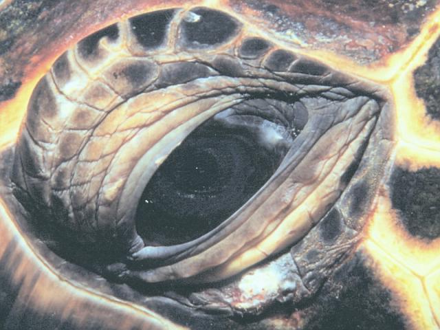
These results posed a dilemma. Although we have images of eyes that are free of tumors, FP is so prevalent at Honokowai that none of them could be used as a standard normal eye. Fortunately, co-author George Balazs provided photographs to use for this purpose.
 |
||
Using these eyes as a baseline, the Honokowai images could be examined for departures from a normal eye. We were able to document both the pre-eruptive stage of FP and the stage of regression that we call "scarred", in which the tumor has disappeared.
 |
||
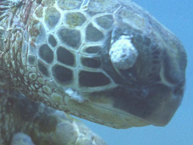
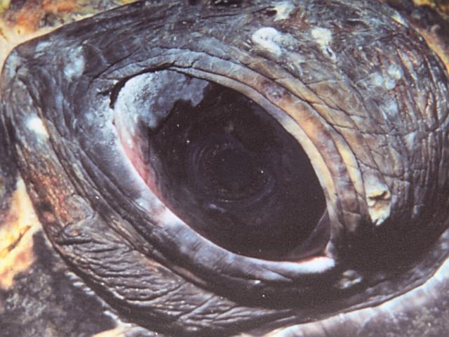
In our experience, the white discolouration along the leading edge advances to swelling, and discolouration on the conjunctiva also expands. Eventually, these abnormalities turn into protuberances that manifest as tumors, as was the case in this example. While it can't be proven that these signs always indicate the onset of FP, every turtle that was documented with this condition has developed ocular tumors.
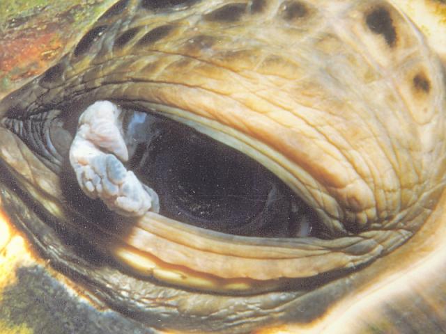
Tumors almost always first erupted in the posterior of the eye. If both corners erupted simultaneously, the posterior tumor was usually the larger. As tumors progress, they appear to be primarily white or pinkish in our images, with a fibrous, wart-like surface, and a polypoid or peduncular form, consistent with the histologic description of ocular tumors prepared by Brooks et al. (1994)
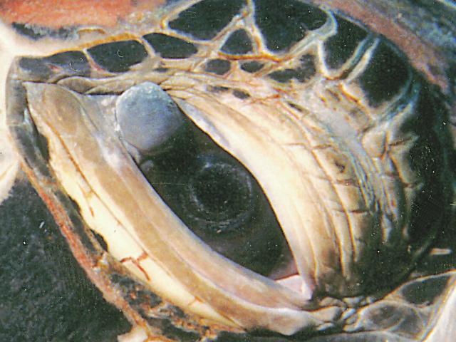
If the animal is lucky, the eye tumors will eventually start to regress. The size stabilizes and starts to decrease. The colour takes on shades of grey, which darken with time. The surface becomes smoother. Eventually, the tumor recedes and darkens so much that it is hard to observe from a distance. This is the condition we refer to as a "black pearl."
Eye tumors usually grow and persist for years, but for four lucky turtles, a remarkable thing happened: the tumors regressed almost immediately. The cycle, from onset to advanced regression, took about a year, demonstrating the possibility of a low level, quickly regressing course for the disease.
 |
||
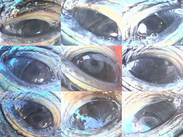
This lack of pleating is seen in every regression case for which we have macro photos. Unfortunately, the history of every animal with this anomaly isn't known, hence the term "scarred" rather than "regressed." It is important to note here, however, that most turtles with eyes classified as scarred also have body tumors that look like they are regressing.
Until now, pre-eruptive or scarred eyes would not be scored as FP. The relationship of these stages to FP was not demonstrated. We believe that we have documented these relationships, however. Certainly, other conditions might account for these anomalies, but in all cases for which there is sufficient history, we can show that the anomalies called "pre-eruptive" do lead to tumors, and the anomalies called "scarred" did result from regression.
It follows that FP is underreported. A normal catch-severity score-release program conducted at Honokowai would inevitably include turtles with no tumors but with pre-eruptive eyes. We believe that these turtles will almost certainly get FP, since we have yet to find an exception.
Additionally, the sample would also include turtles that have had FP and regressed to the point where tumors are no longer visible. We know this because we have documented the history of FP in many of these animals, and Honokowai is home to a number of completely recovered turtles. (Bennett et al. 2000)
 |
||
The records show that four of the turtles without visible tumors are actually regression cases who had ocular tumors. They also show that two of the animals have pre-eruptive eyes and almost certainly will have tumors by next summer. The depth of FP shoots up to 78% (n=14), all of them with some involvement of the eyes. These additional insights give a much clearer picture of the dynamics of FP at Honokowai.
To get an idea about how useful the Honokowai observations might be, we looked at the tumor severity scores for 100 tumored turtles from long-term research conducted in Kaneohe Bay, Oahu, Hawaii (Balazs et al. 2000) during the years 1998-2000. 87% (n=87) of these animals, predominantly juveniles and sub-adults by size, had ocular tumors.
For comparison, all FP-afflicted juvenile and sub-adult turtles (by size) from the years 1998-2000 were selected from the Honokowai database. This yielded 111 turtles. 91% (n=101) of these animals had ocular tumors. Five of the remaining turtles were regression cases, confirmed by photos or videotape, and five fell into our scarred category. This strongly suggests that at least some of the 13 Kaneohe turtles without ocular tumors are regression cases.
The similarities between these Honokowai and Kaneohe Bay data lead us to believe that, yes, the experience gained at Honokowai can be useful when assessing other sites, at least in the Hawaiian archipelago.
From our observations, we have derived some basic lessons. We offer them here in the hope that they might have value to a researcher attempting to assess the scope and magnitude of FP at another site.
The first lesson of Honokowai is:
Examine the eyes.
White discolouration and swelling in the posterior of the eye are the forerunners of FP. A scarred conjunctiva without the pleats characteristic of a normal eye is a sign of FP regression-the "FP signature". Eye examinations are important because body tumors often regress completely and leave no trace, but this is not true for ocular tumors.
The second Honokowai lesson is:
Progressing tumors can be distinguished from regressing tumors.
Graying, smooth, regressing tumors are visually different from "hot" progressing ones--most of which appear an angry white underwater, with a notably different rough, fibrous, or papilloform texture.
|
|
||||||||||||||
| Progressing eye tumor | Regressing eye tumor | ||||||||||||||
This leads to the third lesson:
A turtle with body tumors but no eye tumors is almost certainly a regression case.
In our 245 turtles with FP, just eight were never seen with eye tumors. Two of these turtles were confirmed regression cases. Six of the turtles had eye anomalies consistent with regression, and all eight had body tumors that appeared to be regressing.
Lesson four:
Eye examinations give a better understanding of an individual's FP status.
If a turtle has tumors, it is possible to determine whether they are active or retreating. If tumors are completely absent, normal eyes suggest an animal has never had the disease. Black pearls or an FP signature indicate that it has battled the disease and survived. Pre-eruptive eyes suggest a battle that's yet to be fought.
Lesson five:
Eye examinations give a better approximation of the actual FP dynamics for an area.
This is particularly relevant in Hawaii, because honu demonstrate strong site fidelity. (Bennett et al. In press) The number of turtles who have fought FP successfully--regression cases--will eventually mount up in an area while those who lose the struggle die. It seems likely that the greater the percentage of regression cases, the longer the area has hosted the FP contagion. Similarly, an area that includes primarily tumored turtles and tumor-free turtles with normal eyes strongly suggests that the disease is newly arisen, probably within the previous five years.
 |
||
Examination of the eyes therefore provides a more accurate assessment of FP status in an ohana of Hawaiian green turtles. In comparisons between Honokowai and other Hawaiian sites, there is enough agreement to convince us that our lessons can be useful at those sites.
An old English proverb reads, "The eyes are the window of the soul." We don't know whether sea turtles have souls, but we do think that the FP history of the honu is writ large in their eyes. The tragedy is that the story of erupting FP is read almost exclusively in the eyes of the juveniles. On a positive note, our observations suggest that the eyes provide insight, not just into an individual's FP status, but also into the way FP works its Evil on an entire population of honu. Can this narrative be found in the eyes of non-Hawaiian turtles?
We implore you to look.
We would like to acknowledge Dr. Bob Morris of the Makai Animal Clinic, Kailua, Hawaii, and Dr. Jeanette Wyneken, Florida Atlantic University, for their help in preparing this presentation.
Bennett, P. A., U. Keuper-Bennett, and G. H. Balazs. 2000. Photographic evidence for the regression of fibropapillomas afflicting green turtles at Honokowai, Maui, in the Hawaiian Islands. In Proceedings of the Nineteenth Annual Symposium on Sea Turtle Biology and Conservation, March 2-6, 1999, South Padre Island, Texas, p. 37-39. U.S. Dep. Commer. NOAA Tech. Memo. NMFS-SEFSC-443.
Bennett, P., U. Keuper-Bennett, and G. H. Balazs. In Press. Remigration and residency of Hawaiian green turtles in coastal waters of Honokowai, West Maui, Hawaii. In Proceedings of the Twentieth Annual Symposium on Sea Turtle Biology and Conservation, February 29 - March 4, 2000, Orlando, Florida.
Balazs, G. H. and S. G. Pooley (eds.). 1991. Research plan for marine turtle fibropapilloma. U.S. Dep. Commer., NOAA Tech. Memo. NMFS-SWFSC-156, 113 p.
Work, T. M. and G. H. Balazs. 1999. Relating tumor score to hematology in green turtles with fibropapillomatosis in Hawaii. J. Wildl. Dis. 35(4):804-807.
Brooks, D. E., P. E. Ginn, T. R. Miller, L. Bramson, and E. R. Jacobson. 1994. Ocular fibropapillomas of green turtles (Chelonia mydas). Vet. Pathol. 31:335-339.
Balazs, G. H., S. K. K. Murakawa, D. M. Ellis, and A. A. Aguirre. 2000. Manifestation of fibropapillomatosis and rates of growth of green turtles at Kaneohe Bay in the Hawaiian Islands. In F. A. Abreu-Grobois, R. Briseńo-Dueńas, R. Márquez-Millán, and L. Sarti-Martínez (comps.), Proceedings of the Eighteenth International Sea Turtle Symposium, March 3-7, 1998, Mazatlán, Sinaloa, México, p. 112-113. U.S. Dep. Commer. NOAA Tech. Memo. NMFS-SEFSC-436.
 The Sickbay
 Table of Contents