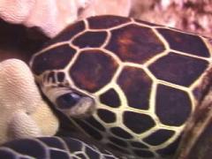
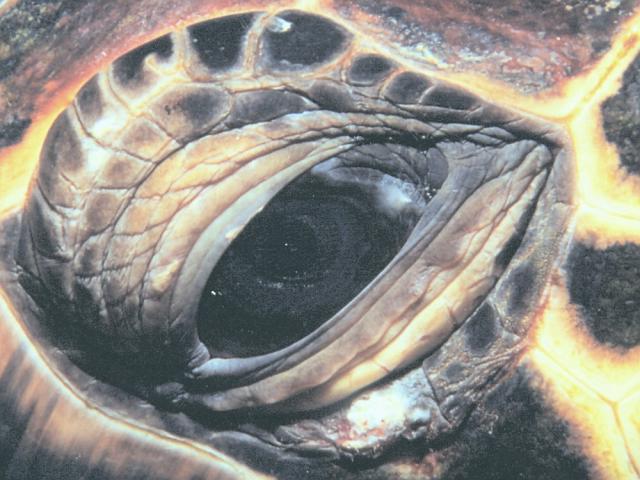
These photographs were taken underwater at Honokowai by Ursula Keuper-Bennett. They illustrate pre-eruptive eyes. Eyes signal problems well before a tumour erupts. In our experience, ocular anomalies associated with FP can be detected typically a full year before a lump develops.
Note the white discolouration and spotting on the posterior conjunctiva. These are the early signs of fibropapilloma. Note also that the conjunctiva in these examples still shows the pleating of a normal eye.
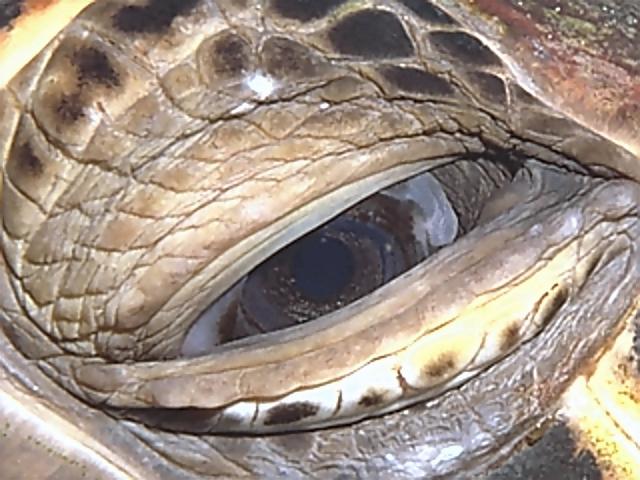
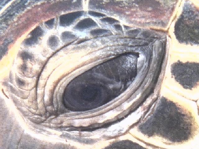
This is Limu in 1995. This is the most subtle ocular anomaly we've recorded to date. There is the faintest hint of white showing on the leading edge of the posterior conjunctiva. A barely detectable "hotspot" extends from the leading edge into the cornea of the eye.
|
A year later, a small lump had grown from the 1995 hotspot 56K JPEG |
|||||||
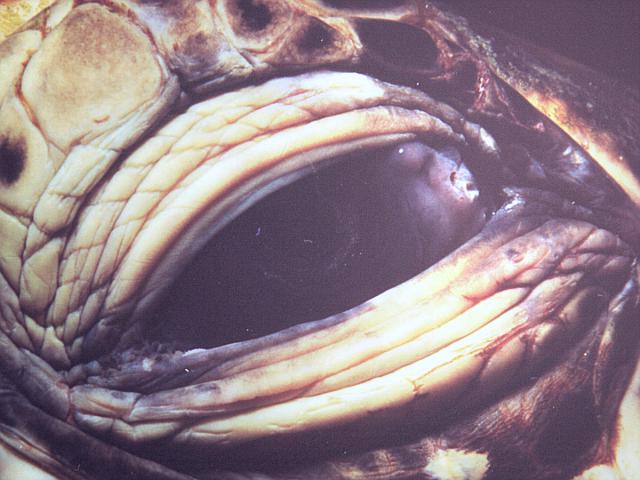
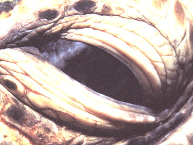
While the tumors in Limu's right eye have grown significantly in size by Summer 2000, the tumor in the left eye has remained small. This might have something to do with the tumor's site of origin--corneal rather than conjunctival.
This tumor is corneal rather than conjunctival 83K JPEG |
|
|||||||
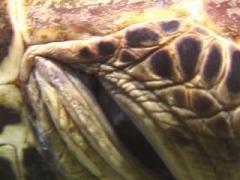
Turtle 2000-55, showing two obvious hotspots on the leading edge of the posterior conjunctiva. Whitening at the anterior corner of the eye hints at potential problems also. Should this turtle be around for Summer 2001 we expect tumours both front and back, with the posterior being larger.
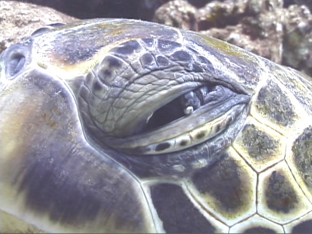
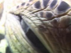
2000-47's right eye shows two unmistakeable signs of pre-eruptive FP. The leading edge of the posterior conjunctiva shows white discolouration and swelling (lifting away from the cornea). Should Turtle 2000-47 be around in Summer 2001 we expect this right eye to have at least one tumour originating from the conjunctiva.
|
Several small white lumps buckle the conjunctiva of 2000-47's left eye 48K JPEG |
|||||||
2000-47's left eye was in the early eruptive stage in 2000, with several small white lumps buckling the conjunctiva. It is not unusual for one eye to be ahead of the other in terms of tumour development. For example, a turtle can have a size C (fist-sized) tumour in one eye while the other eye may host a tumour no bigger than a pea! We call this asymmetrical ocular FP.
Video often is more revealing than a still photograph because the motion gives the image more form and depth. Click on the still images below to see the video clips.
| We first sighted Akebono in 1999. The little honu was about 38-40 CCL--tiny--so we named her after the Sumo Grand Master. It was our hope that Akebono would grow up to be *B*I*G*. Unfortunately both eyes showed the early harbingers of fibropapilloma disease. The left showed a white discolouration running along the leading edge of the posterior conjunctiva. This ocular anomoly is a common sign of the pre-eruptive stage of this disease. |
|
|||||||
|
This is what Akebono's left eye looked like a year later--summer 2000. The conjunctiva is white, swollen and a tumour "hotspot" threatens. We fully expect a true tumor in Akebono's eye for Summer 2001. | |||||||
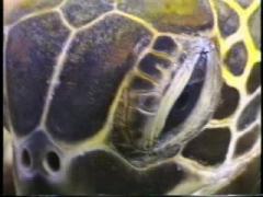
| This is turtle 2000-94. The right eye shows white discolouration along the leading edge of the posterior conjunctiva. There are two "hotspots" on this band of white that will likely erupt into tumors. |
|
|||||||
|
This video of turtle 2000-94's right eye was shot on the same dive--the honu opens his eye wider and reveals two true tumours. We have listed the turtle as "FP progressive, year 1." | |||||||
|
|
The eyes have it | ||||
|
|
The Sickbay | ||||
|
|
Table of Contents | ||||